C-Path (the accredited histopathology laboratory within SAS Global) has expanded its portfolio and capabilities by introducing state-of-the-art veterinary histopathology services to support poultry, swine, ruminant and companion animal diagnostic histopathology. Founded in 2021 and joined SAS Global in 2025, C-Path allows CRO’s, biotechs, pharmaceutical companies, and academic organizations to focus on understanding the complexities of their research and less on the operations.
C-Path offers end-to-end histopathology assessments and a flexible workflow, allowing customers to outsource exactly the level of service and support they require. This workflow is built with cost-effective and reliable channels that are digitized, automated, and efficient, such as the TxConnect track-and-trace ordering system combined with AI assessment tools.
Poulpharm
A leading veterinary diagnostic and contract research organization with branches in Asia, Europe and Latin America, and also member of SAS Global.
Veterinary diagnostic pathology is the backbone of veterinary diagnostics and is crucial for accurately diagnosing animal diseases, guiding treatment and prognosis, and advancing biomedical research by examining tissues and cells for disease clues, bridging the gap between clinical signs and underlying causes for better animal welfare and health.
For several diseases/conditions, diagnostic histopathology is a must unreplaceable endpoint to determine differential diagnoses or confirm final diagnosis.
Examples of these diseases or conditions that cannot be diagnosed without histopathology include but are not limited to feline and canine tumors, feline and canine skin diseases, companion animal surgical pathology (biopsy services) and most of infectious diseases of multiple species (e.g. Swine proliferative enteropathy, Avian mycoplasmas, Mycobacterial diseases, Canine parvovirus, Feline infectious peritonitis (FIP), etc)
In order to support Poulpharm diagnostic work, C-Path took the strategic decision to expand its service to also include multispecies diagnostic histopathology as follows:
Swine, Ruminant and Poultry diagnostics
-Full necropsy and macroscopic examination performed on the farm by our experienced SAS veterinarians and discussed with our board certified veterinary pathologists. Representative specimens will be collected from affected organs and sent to C-Path to be processed for histopathology by experienced histotechnologists.
-Complete and timely histopathology assessment by highly experienced board certified veterinary diagnostic pathologists who will generate a full report containing descriptions of findings, differential diagnosis and whenever possible final diagnosis/interpretation and/or suggestions for further testing/staining.
-In certain cases and upon discretion of the examining pathologists, special specific stains for suspected infectious diseases can be performed to confirm the diagnosis. Example of these stains include Gram stain (Fig.1) for bacteria, Ziehl Nelsen stain for Mycobacteria spp, Warthin starry stain for higher bacteria, Brachyspira spp and Lawsonia intracellularis (Fig.2), and immunohistochemistry for Mycoplasma spp, and selected viral diseases (e.g. PVC2, ILT and PRRS).
Companion animal diagnostics
-Complete histopathologic analysis for canine and feline surgical biopsies by highly experienced board certified surgical veterinary pathologists with a fast turnaround time from picking up specimens to full reporting (up to maximum of 72 hrs)
-Extended list of special stains and immunostains (IHC or IF) for different tumors (tumor markers) and infectious diseases. Examples include same stains mentioned in the swine and poultry diagnostics, IHC for Canine Parvovirus, FIP, and more.
-Cytology services including smears from tissue imprint and fine needle aspirates cytology.
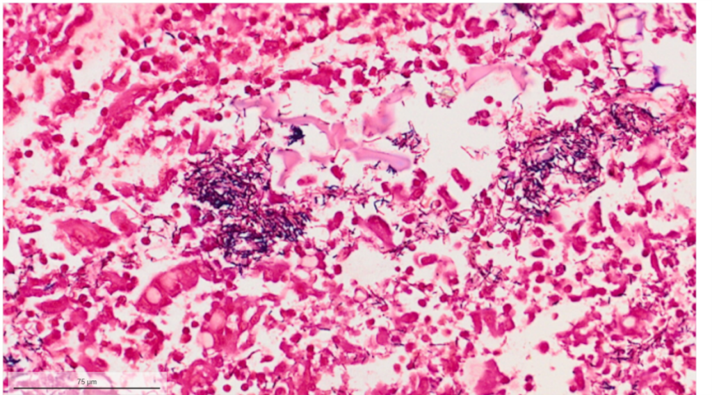
Fig.1 Gram stain of swine colon section reveals gram-positive bacilli (arrows) reminiscent of Clostridial spp.
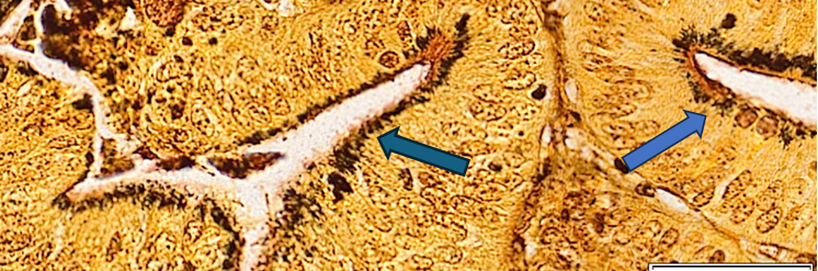
Fig. 2: Warthin Starry silver stain of swine colon affected with L. intracellularis reveal numerous argyrophilic curved small bacilli (arrows) concentrated in the apical cytoplasm of hyperplastic mucosal epithelial cells.
Recent Comments